Rapidly decreasing Prolactin result

HOSP # WARD Endocrinology Clinic
CONSULTANT John Stanfliet   DOB/AGE 36 y Female

Abnormal Result

A low prolactin result was obtained in a patient in whom a macroadenoma was suspected:

Prolactin: 1.3 mIU/L

Presenting Complaint

The patient presented with headache and decreased visual acuity (more specifically peripherally).

History

There were bilateral galactorrhoea, amenorrhoea, and as noted above, headache and visual disturbances.

The patient had received Cabergoline (a dopamine receptor agonist on D2 receptors) for the past 4 months.

Examination

As above

Laboratory Investigations

Date Prolactin (mIU/L)
02/2019 106 (Recovery of 80% following PEG precipitation)
05/2019 135
06/2019 85
08/2019 1.3 (1.59 with a 1:10 dilution; 3.94 with a 1:50 dilution)
Prolactin Results

Other Investigations

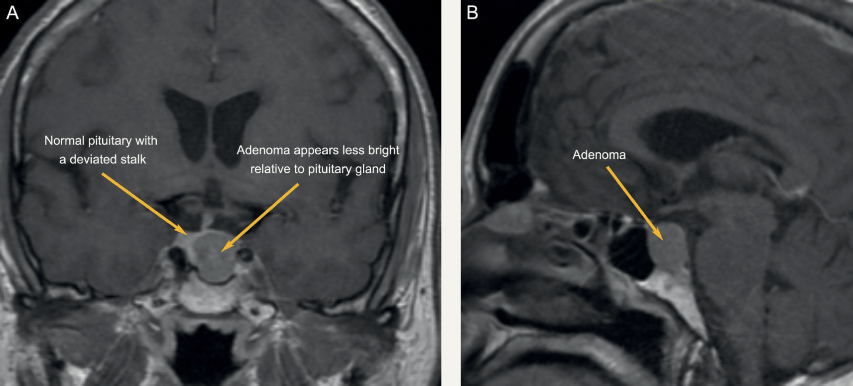
MRI Head was booked for the following week. Interestingly, even in prolactin secreting tumours, the correlation between tumour size and prolactin level is limited. MRI head remains a vital investigation.

Final Diagnosis

An Introduction to Sellar Masses (Chapter 10) - Clinical Neuroendocrinology
Pituitary Macroadenoma

Take Home Message

During pregnancy the concentration of prolactin rises under the influence of elevated estrogen and progesterone production. The stimulating action of prolactin on the mammary gland leads post partum to lactation. Hyperprolactinemia (in men and women) is the main cause of fertility disorders. The determination of prolactin is utilized in the diagnosis of anovular cycles, hyperprolactinemic amenorrhea and galactorrhea, gynecomastia and azoo-spermia. Prolactin is also determined when breast cancer and pituitary tumors are suspected. As in this case, a pituitary tumour was suspected, hence the repeated prolactin results.

As was noted in another short case, our assay on the Roche platform does measure all forms of prolactin, and when a high result is obtained (above the gender-specific reference range) it is recommended to measure the recovery after PEG precipitation.

Figure 1

Dr. John Stanfliet (pathologist at Pathcare) repied to the above case with very valuable comments:

  • We use Beckman Coulter DxI, an immunoassay that is not affected by macroprolactin (I’ve include an article that shows this).
  • Even in prolactin secreting tumours, the correlation between tumour size and prolactin level is limited.  MRI head remains a vital investigation.
  • Some prolactin secreting tumours also secrete other pituitary hormones such as growth hormone.
  • I would ascribe the reduction in PRL to the Carbegoline and wonder whether the dose has been increased.
  • Dr. Pete Berman would often suggest a mixing study: find a sample with high PRL, mix it 50/50 with this sample, and measure it to see whether there is some interferant in this sample. 


Iron deficiency anemia with a twist

HOSP # MRN53499748 WARD C15 Emergency Unit
CONSULTANT   Dr. Heleen Vreede DOB/AGE 16 y/o Female

Abnormal Result

TSH: 42.9 mIU/L (0.51 – 4.30)

Free T4: 7.1 pmol/L (12.6 – 21.0)

Presenting Complaint

Patient presented with signs and symptoms of iron deficiency anemia.

History

The patient is a known hemophilia B carrier (Factor IX deficiency or Christmas disease) with menorrhagia and accompanying iron deficiency anemia.

Examination

The patient presented to the emergency rooms with symptoms of severe weakness and had occasional severe menorrhagia.

Unfortunately the physical examination details are not available.

Laboratory Investigations

  • Iron: 4.5 umol/L (9-30.4) Low
  • Transferrin 3.92 g/L (2-3.6) High
  • % Saturation 5% (15 -50) Low
  • Ferritin 8 ug/L (13- 68) Low

Other Investigations

Anti-Thyroglobulin (anti-TG) antibodies as well as Anti-Thyroid Peroxidase (anti-TPO) antibodies were positive in this patient.

These antibodies was suggested after the results of the TSH and reflexed free-T4 became available and hence after-requested.

Final Diagnosis

The pattern of significantly raised TSH with the significantly low free-T4 and the raised anti-TPO and anti-Thyroglobulin antibodies suggest primary hypothyroidism.

Take Home Message

It’s always worth doing a TSH to screen for thyroid disease when a patient presents with weakness / tiredness, irrespective of the age.

Primary hypothyroidism due to an auto-immune mediated destruction of the thyroid gland tissue is the most common hypothyroid condition and is confirmed by measuring the common anti-thyroid antibodies: Anti-TPO and Anti-TG antibodies. There is likely not much indication for performing these antibodies more than once after diagnosis of hypothyroidism and some clinicians argue it not necessary to even perform these antibody measurements.

Congenital hypothyroidism is one of the congenital disorders causing cretinism which is most preventable by newborn screening. Even though not likely congenital in this patient, it’s worth considering on the differential diagnosis.

The patient was on iron supplements as well as Factor IX injections. I was not aware of an association between Factor IX deficiency and Hypothyroidism, but my Haematology colleagues across the corridor told me the following:

Factor IX deficiency is an X-linked recessive disorder. This makes it unlikely for a female to get this disease. Auto-immune diseases is much more likely in females. There is also a form of Christmas disease where one produces antibodies to factor IX, which yields it inactive, hence presenting as Factor IX deficiency.

This, although unlikely, presents an interesting thought for this unusual presentation in this 16-year old female. Acquired deficiencies of most clotting factors have been described.

However, upon discussion with the attending clinician it seems that the patient did have a clear family history of Christmas disease, hence the presentation.