Query EDTA contamination

HOSP # MRN96038757 WARD F17 Surgical ward
CONSULTANT   Dr. Jody Rusch DOB/AGE 26 y male

Abnormal Result

Potassium more than 10 mmol/L on the ion-selective electrode.

Presenting Complaint

The patient was admitted in surgery after bowel surgery, on total parenteral nutrition.

History

Surgery was done due to bowel obstruction.

Examination

Not available.

Typically:

A hallmark of small bowel obstruction is dehydration, which manifests as tachycardia, orthostatic hypotension, and reduced urine output, and, if severe, dry mucus membranes.

Abdominal inspection will identify a variable degree of abdominal distention.

Abdominal auscultation – Acute mechanical bowel obstruction is characterized by high-pitched “tinkling” sounds associated with the pain. With significant bowel distention, bowel sounds may become muffled, and as the bowel progressively distends, bowel sounds can become hypoactive.

Abdominal percussion – Distention of the bowel results in hyperresonance or tympany to percussion throughout the abdomen. However, fluid-filled loops will result in dullness. If percussion over the liver is tympanitic rather than dull, it may be indicative of free intra-abdominal air. Tenderness to light percussion suggests peritonitis.

Abdominal palpation may identify any abdominal wall or groin hernias, or abnormal masses.

Laboratory Investigations

Date 16/03/2020 14/03/2020 13/03/2020 12/03/2020 12/03/2020 11/03/2020 10/03/2020 08/03/2020
Time 07:22 10:38 10:30 19:59 12:32 21:23 16:05 12:53
Na 144   141     141          δ-  138     143   δ+  145     140  
K >10 2.6   3.0 L   3.0 L INVH   3,2 L δ+  3,7     3.0 L
Cl                                                        
Urea 4.4   5,1     5.0            5,5     6,9   δ+  5,5     2,5  
Creat 47    60 L    60 L           64      67      66      62 L
Ca 1.63  2,24    2,27           2,13 L  2,23    2.30    2,23  
Mg 0.38 δ- 0.67   δ+ 0.90           0.77    0.82    0.75    0.74  
Phos 1.1  1.50 H  1,31           1,28    1,38    1,23    1,17  
Uric acid                                                        
Total prot CEGK                      CEGK                     
Alb 30                         39                    40  
Total bili <3                          3 L                   4 L
Conj bili 2                      INVH                   2  
ALT 35                         28                    32  
AST 33                      INVH                  28  
ALP 118                        137 H                 158 H
GGT 99                         96 H                 113 H
LD 138                      δ+  465 H                 317 H
CRP 2                          7                     6  
Table 1 – Results in bold indicative of likely EDTA contamination.

Other Investigations

Repeated results later in the afternoon:

Date 16/03/2020 16/03/2020
Time 13:04 07:22
Na 144 144
K δ+  3,3 L >10
Cl              
Urea 4.6 4.4
Creat 55 47
Ca 2.2 1.63
Mg 0.56 0.38
Phos 1.06 1.1
Uric acid              
Total prot CEGK CEGK
Alb 37 30
Total bili     3 L <3
Conj bili 2 2
ALT 46 35
AST 40 33
ALP 151 118
GGT 121 99
LD 230 138
CRP 2 2
Initial results on the right. Repeated (new) results on the left.

Final Diagnosis

Likely EDTA contamination causing a falsely elevated potassium, decreased Calcium, Magnesium and ALP. The clinician was contacted and it was indeed medical undergraduate students who had taken the bloods, probably not realizing the order of draw, or toppling up the serum blood with some of the blood taken in an EDTA tube. This is evidenced by the high potassium, low calcium, magnesium and ALP. It is however evident that most other analytes were also lower than the repeat bloods later that day, hence:

Another likely possibility of the results in question could have been drip line contamination due to a potassium-containing fluid. The patient was indeed on total par-enteral nutrition, which usually contain large doses of potassium. This could be explained by the dilution of most analytes (as opposed to the raised potassium and normal sodium).

Take Home Message

It does not require much potassium EDTA contamination to evoke spuriously abnormal results. Potassium EDTA works as an anticoagulant by inhibiting clotting by chelation of the divalent cations such as calcium and magnesium, essential for the divalent cation-dependent proteolytic enzymes involved in the clotting cascade.

Gross potassium EDTA contamination of blood samples can be recognized by unexpected marked pseudohyperkalaemia and pseudohypocalcaemia. Serum alkaline phosphatase (ALP) activity can also be reduced in the presence of potassium EDTA contamination. Additionally, aspartate transaminase, alanine transaminase, lactate dehydrogenase, creatine kinase, amylase, unsaturated iron-binding capacity and bicarbonate can all be detrimentally affected in the presence of potassium EDTA contamination. Notably, some papers report potassium EDTA contaminated samples were mainly from inpatients compared to outpatients and primary care and the authors speculated that this is because blood samples in outpatients and general practice are largely but not exclusively collected by trained phlebotomists. It is our job as laboratorians to educate the newly trained clinicians about order of draw.

It is unfortunate that I couldn’t locate the undergraduate student who had taken these bloods, but at least the attending clinician was made aware of EDTA contamination.


Discrepant TFT’s

HOSP # WARD Endocrine Clinic (OPD)
CONSULTANT  Dr. Jody Rusch DOB/AGE 32 Year female

Abnormal Result

The clinician, an endocrinologist, phoned about discrepant results: Suppressed TSH, Low Free T4 and Normal (upper end of reference interval) Free T3.

Date 09/12/2020
TSH (mIU/L)  0.05 L
Free T4 (pmol/L)   4,8 L
Free T3 (pmol/L)   6,4  

Presenting Complaint

The patient was known with Graves Disease complicated by quite severe Graves Eye Disease (orbitopathy).

History

Known with Graves disease with positive antibodies to TSH-receptors.

Examination

The clinical examination for this patient is not available, but the following is important:

Interestingly, patients may have no ocular symptoms at all, but may sometimes be distressed by the appearance of their eyes. The major ocular symptoms include:

  • A gritty or foreign object sensation
  • Excessive tearing that is often made worse by exposure to cold air, wind, or bright lights
  • Eye or retroocular discomfort or pain
  • Blurring of vision
  • Diplopia
  • Color vision desaturation
  • Loss of vision in severe cases

The characteristic signs of Graves’ orbitopathy are proptosis (exophthalmos), tearing, and periorbital edema. In more severe disease, there may be severe conjunctival inflammation and ulceration from over exposure.

Laboratory Investigations

Date 09/12/2020 11/05/2020 08/11/2019 24/05/2019 29/01/2019 10/12/2018
TSH (mIU/L)  0.05 L (Rerun 0.05)  0.02 L <0.01 L         <.01 L  <.01 L
Free T4 (pmol/L)   4,8 L (Rerun 4.9)  65,7 H δ+>100.0 H  48,4 H  42,7 H    44 H
Free T3 (pmol/L)   6,4  (Rerun 6.4)                       19,6 H       

As above, the history of Graves disease is clear, which includes a suppressed TSH and raised Free T4 and Free T3.

Other Investigations

The Free T4, Free T3 and TSH was re-run on 10/12/2020, QC checked on these three analytes (all was within normal range) and pre-analytical labeling errors excluded as far we could.

Final Diagnosis

Graves eye disease, now with hypothyroidism.

Take Home Message

In Graves’ disease, the main auto-antigen is the thyroid-stimulating hormone (TSH) receptor (TSHR), which is expressed primarily in the thyroid but is also expressed in adipocytes, fibroblasts, and a variety of additional sites and appears to be closely aligned with the insulin-like growth factor 1 (IGF-1) receptor. TSHR antibodies and activated T cells also play an important role in the pathogenesis of Graves’ orbitopathy by activating retro-ocular fibroblast and adipocyte TSHR and IGF-1 receptors and initiating a retro-orbital inflammatory environment.

The retro-orbital tissue (and ocular muscles) increase in volume due to this inflammatory milieu, fibroblast proliferation and the accumulation of hydrophilic glycosaminoglycans (GAG’s), most notably hyaluronic acid.

Sometimes orbitopathy occurs in patients with hypothyroidism (high TSH, low free T4) due to classical chronic autoimmune thyroiditis (Hashimoto’s disease), and these patients may have stimulating TSH receptor (TSHR) antibodies but inadequate thyroid reserve.

In summary, the most important factors for development of Graves Eye Disease (orbitopathy) seems to be:

  • Graves Orbitopathy antigen (which is the TSH-receptor): these are expressed extra-thyroidally, especially retro-orbitally.
  • Role of TSH receptor antibodies
  • Role of T-cells: Retroocular fibroblasts secrete GAG in response to cytokines such as interferon gamma and tumor necrosis factor (TNF)-alpha secreted by helper (CD4+) T cells of the Th1 type.

In cases of hypothyroidism, the action of deiodinase is increased to protect against the effects of hypothyroidism, likely the explanation of the increased Free T3 in this patient (compared to the low Free T4).


Rapidly decreasing Prolactin result

HOSP # WARD Endocrinology Clinic
CONSULTANT John Stanfliet   DOB/AGE 36 y Female

Abnormal Result

A low prolactin result was obtained in a patient in whom a macroadenoma was suspected:

Prolactin: 1.3 mIU/L

Presenting Complaint

The patient presented with headache and decreased visual acuity (more specifically peripherally).

History

There were bilateral galactorrhoea, amenorrhoea, and as noted above, headache and visual disturbances.

The patient had received Cabergoline (a dopamine receptor agonist on D2 receptors) for the past 4 months.

Examination

As above

Laboratory Investigations

Date Prolactin (mIU/L)
02/2019 106 (Recovery of 80% following PEG precipitation)
05/2019 135
06/2019 85
08/2019 1.3 (1.59 with a 1:10 dilution; 3.94 with a 1:50 dilution)
Prolactin Results

Other Investigations

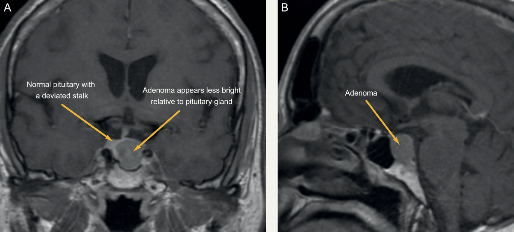
MRI Head was booked for the following week. Interestingly, even in prolactin secreting tumours, the correlation between tumour size and prolactin level is limited. MRI head remains a vital investigation.

Final Diagnosis

An Introduction to Sellar Masses (Chapter 10) - Clinical Neuroendocrinology
Pituitary Macroadenoma

Take Home Message

During pregnancy the concentration of prolactin rises under the influence of elevated estrogen and progesterone production. The stimulating action of prolactin on the mammary gland leads post partum to lactation. Hyperprolactinemia (in men and women) is the main cause of fertility disorders. The determination of prolactin is utilized in the diagnosis of anovular cycles, hyperprolactinemic amenorrhea and galactorrhea, gynecomastia and azoo-spermia. Prolactin is also determined when breast cancer and pituitary tumors are suspected. As in this case, a pituitary tumour was suspected, hence the repeated prolactin results.

As was noted in another short case, our assay on the Roche platform does measure all forms of prolactin, and when a high result is obtained (above the gender-specific reference range) it is recommended to measure the recovery after PEG precipitation.

Figure 1

Dr. John Stanfliet (pathologist at Pathcare) repied to the above case with very valuable comments:

  • We use Beckman Coulter DxI, an immunoassay that is not affected by macroprolactin (I’ve include an article that shows this).
  • Even in prolactin secreting tumours, the correlation between tumour size and prolactin level is limited.  MRI head remains a vital investigation.
  • Some prolactin secreting tumours also secrete other pituitary hormones such as growth hormone.
  • I would ascribe the reduction in PRL to the Carbegoline and wonder whether the dose has been increased.
  • Dr. Pete Berman would often suggest a mixing study: find a sample with high PRL, mix it 50/50 with this sample, and measure it to see whether there is some interferant in this sample. 


Iron deficiency anemia with a twist

HOSP # MRN53499748 WARD C15 Emergency Unit
CONSULTANT   Dr. Heleen Vreede DOB/AGE 16 y/o Female

Abnormal Result

TSH: 42.9 mIU/L (0.51 – 4.30)

Free T4: 7.1 pmol/L (12.6 – 21.0)

Presenting Complaint

Patient presented with signs and symptoms of iron deficiency anemia.

History

The patient is a known hemophilia B carrier (Factor IX deficiency or Christmas disease) with menorrhagia and accompanying iron deficiency anemia.

Examination

The patient presented to the emergency rooms with symptoms of severe weakness and had occasional severe menorrhagia.

Unfortunately the physical examination details are not available.

Laboratory Investigations

  • Iron: 4.5 umol/L (9-30.4) Low
  • Transferrin 3.92 g/L (2-3.6) High
  • % Saturation 5% (15 -50) Low
  • Ferritin 8 ug/L (13- 68) Low

Other Investigations

Anti-Thyroglobulin (anti-TG) antibodies as well as Anti-Thyroid Peroxidase (anti-TPO) antibodies were positive in this patient.

These antibodies was suggested after the results of the TSH and reflexed free-T4 became available and hence after-requested.

Final Diagnosis

The pattern of significantly raised TSH with the significantly low free-T4 and the raised anti-TPO and anti-Thyroglobulin antibodies suggest primary hypothyroidism.

Take Home Message

It’s always worth doing a TSH to screen for thyroid disease when a patient presents with weakness / tiredness, irrespective of the age.

Primary hypothyroidism due to an auto-immune mediated destruction of the thyroid gland tissue is the most common hypothyroid condition and is confirmed by measuring the common anti-thyroid antibodies: Anti-TPO and Anti-TG antibodies. There is likely not much indication for performing these antibodies more than once after diagnosis of hypothyroidism and some clinicians argue it not necessary to even perform these antibody measurements.

Congenital hypothyroidism is one of the congenital disorders causing cretinism which is most preventable by newborn screening. Even though not likely congenital in this patient, it’s worth considering on the differential diagnosis.

The patient was on iron supplements as well as Factor IX injections. I was not aware of an association between Factor IX deficiency and Hypothyroidism, but my Haematology colleagues across the corridor told me the following:

Factor IX deficiency is an X-linked recessive disorder. This makes it unlikely for a female to get this disease. Auto-immune diseases is much more likely in females. There is also a form of Christmas disease where one produces antibodies to factor IX, which yields it inactive, hence presenting as Factor IX deficiency.

This, although unlikely, presents an interesting thought for this unusual presentation in this 16-year old female. Acquired deficiencies of most clotting factors have been described.

However, upon discussion with the attending clinician it seems that the patient did have a clear family history of Christmas disease, hence the presentation.