Bicarbonate HCO3 CO2 CO3

Method

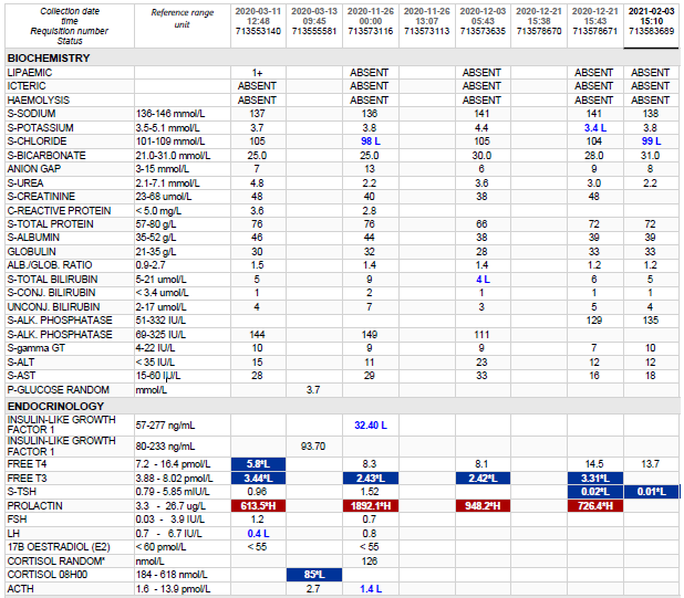
Bicarbonate is the second largest fraction of the anions in plasma. Included in this fraction are the bicarbonate (HCO3) and carbonate (CO32-) ions, as well as the carbamino compounds. At the physiological pH of blood, the concentration of carbonate is 1/1000 that of bicarbonate. The carbamino compounds are also present in such low quantities that they are generally not mentioned specifically.
Several different methods for the determination of bicarbonate in serum and plasma have been reported. Most of these procedures utilize acidification of the sample and conversion of all carbon dioxide forms to CO2 gas. The amount of gas formed is measured by manometric or volumetric devices, ion selective electrodes, or spectrophotometric techniques. These methods are either cumbersome, time-consuming, technique-oriented, and/or require special equipment.
Enzymatic procedures using phosphoenolpyruvate carboxylase (PEPC) have been described.
The bicarbonate content of serum or plasma is a significant indicator of electrolyte dispersion and anion deficit. Together with pH determination, bicarbonate measurements are used in the diagnosis and treatment of numerous potentially serious disorders associated with acid-base imbalance in the respiratory and metabolic systems.

Test principle

Bicarbonate reacts with phosphoenolpyruvate (PEP) in the presence of PEPC to produce oxaloacetate and phosphate:

PEPC
PEP + HCO3 —> oxaloacetate + H2PO4
The above reaction is coupled with one involving the transfer of a hydrogen ion from NADH analog to oxaloacetate using MDH.

MDH

Oxaloacetate + NADH analog + H+ —> malate + NAD+ analog
The resultant consumption of NADH analog causes a decrease in absorbance, which is proportional to the concentration of bicarbonate in the sample being assayed.


Raised fecal osmolar gap

HOSP # MRN123486438 WARD Victoria Hospital Pediatric Ward
CONSULTANT   Dr Jody Rusch DOB/AGE 2y male

Abnormal Result

Fecal osmolar gap 152 mmol/L (<100 mmol/L)

Presenting Complaint

The patient presented with chronic diarrhoea upon which fecal chemistry was requested.

History

The full history is not known.

He presented with an increased anion gap metabolic acidosis a week prior to the stool investigations being requested.

The most common explanation thereof is likely two-fold in the acute setting when the child presented:

  • A fasting response with lipolysis and generation of ketone bodies leading to the acidosis
  • Dehydration leading to hypoperfusion of tissues with a lactic acidemia
    • Uremia and the increased creatinine in the child was indicative thereof

Examination

Not available

Laboratory Investigations

Stool chemistry:
Faecal sodium 22 mmol/L
Faecal potassium 39.4 mmol/L
Faecal osmolality 275 mOsm/kg
Faecal osmolarity (calculated) 123 mmol/L
Faecal osmolar gap 152 mmol/L
A faecal osmolar gap (the difference between measured and calculated osmolarity) of > 100 mmol/L suggests the presence of poorly absorbed solutes.

Other Investigations

Stool reducing substances: 1+

If a positive stool reducing substances is found, characterization of the reducing substance follows. In our laboratory, this is done by thin layer chromatography.

Test Item 23/04/2021 (6 days later) 17/04/2021 (Presentation)
Na   138     135 L
K   4,2       4  
Cl   106     102  
Bicarb δ+   20 L    13 L
Anion gap    16      24 H
Urea δ-  2,2     6,8 H
Creat    25      32 H
CRP            4  

Final Diagnosis

Chronic (or persistent) diarrhoea: In this case the stool microscopy also showed species of Cryptosporidium, likely the cause of the prolonged diarrhoea. Also the relative lactase deficiency after episodes of enteritis is common due to the lactase enzyme being primarily located at the apical surface of the microvili of the enterocytes, also the common site of infection of various infective organisms, such as cryptosporidium.

Take Home Message

A faecal osmolar gap (the difference between measured and calculated osmolarity) of >100 mmol/L suggests the presence of poorly absorbed solutes. A “quick and dirty” way of excluding this is by doing a dipstick of fecal fluid. In cases where diarrhea has persisted for more than two weeks, testing the stool for glucose and pH can be helpful in identifying those children with severe villous atrophy. This can be done easily at the bedside with a urine dipstick if available. Glucose test tape, nitrazine paper, and Clinitest tablets also have been used. A stool glucose of greater than 2+ or a pH of less than 5.0 suggests substantial villous atrophy.

A low stool osmotic gap suggests secretory diarrhea, wherein the digestive tract is hyperpermeable and losing electrolytes, while a high gap suggests osmotic diarrhea, wherein the digestive tract is unable to absorb solutes from the chyme, either because the digestive tract is hypopermeable (e.g. inflammation), or non-absorbable compounds (e.g. Epsom Salts, Lactose) are present. The reason for this is that secreted sodium and potassium ions make up a greater percentage of the stool osmolality in secretory diarrhea, whereas in osmotic diarrhea, other molecules such as unabsorbed carbohydrates are more significant contributors to stool osmolality.


Raised fecal calprotectin

HOSP # WARD GIT clinic
CONSULTANT   Dr. Heleen Vreede DOB/AGE 59 y male

Abnormal Result

Faecal calprotectin >6000 ug/g stool

Presenting Complaint

59 y male, presenting with diarrhoea and bloody mucus per rectum

History

This is a 59 year old male known with ulcerative colitis proctitis who now has a suspected flare.

Ulcerative colitis (pancolitis) diagnosed 2009.

Histological history

2017: Mild focal active colitis noted on Histology

2019: Sections of rectal mucosa showed features of active chronic proctitis. The crypts showed distortion with focal areas of crypt abscesses noted. The lamina propria was expanded by reactive polymorphous mature lymphocytes with conspicuous eosinophils.

Examination

Unknown

One would look for especially extra-intestinal manifestations of Ulcerative Colitis

Laboratory Investigations

Histology: Sections of colon demonstrate a severe acute colitis with cryptitis , crypt abscess and numerous neutrophils in the lamina propria on a background of chronic changes illustrated by architectural disarray and glandular atrophy. 

Other Investigations

Apart from the colonoscopy and histology, one needs to evaluate for other autoimmune disorders in the gastro-intestinal tract, especially complications of primary sclerosing cholangitis. No biochemical signs thereof was present.

Test (units) Result
Creat (umol/L)   122 H
MDRD    53  
CKD-EPI    56  
Alb (g/L)    44  
Total bili (umol/L)     4 L
Conj bili (umol/L)     2  
ALT (U/L)    18  
AST (U/L)    30  
ALP (U/L)    77  
GGT (U/L)    16  
CRP (U/L)     2  

Final Diagnosis

Inflammatory Bowel Disease (Ulcerative colitis)

Take Home Message

We have in recent years started to offer this test. One of our recently qualified pathologists, Dr. Justine Cole, was responsible for the method validation of this assay at our laboratory. There were quite a few difficulties with the validation, mainly due to stool being a difficult to work with matrix and sample stability when transported.

In summary:

Faecal calprotectin is excreted in excess into the intestinal lumen during the inflammatory process and so can act as a marker for inflammatory diseases of the lower gastrointestinal tract. Faecal calprotectin testing is recommended in patients with recent onset lower gastrointestinal symptoms, if cancer is NOT suspected, for the
differential diagnosis of inflammatory bowel disease (IBD e.g., Chrohn’s disease, ulcerative colitis) or irritable bowel syndrome (IBS).

Faecal calprotectin <=50 ug/g stool is negative, i.e., supports IBS.

Faecal calprotectin >50 ug/g stool is positive, i.e., supports IBD.


Primary amenorrhoea with ulcerative colitis

HOSP # WARD
CONSULTANT   DOB/AGE 15 y girl

Abnormal Result

This patient was discussed at a combined Endocrinology / Chemical Pathology meeting.

Total bilirubin: 281 umol/L

Presenting Complaint

The patient was a candidate for a liver transplant, but was referred to the endocrinology department for the short stature and primary amenorrhoea prior to surgery.

History

She was diagnosed with ulcerative colitis in 2016 (@ 12y age) and primary sclerosing cholangitis. Breast development started in 2018 (@14 years), but no menstrual cycles started ever since.

She has one younger sister which is well currently at 4 y age.

Birth weight was 3.8 kg.

Medication

Patient was receiving steroids and sulfasalazine intermittently.

For portal hypertension she is also receiving furosemide and spironolactone

Vitamin D supplements are also given

Examination

Height (114cm) for age: <3rd percentile

Weight 35 kg

Breasts well developed – Tanner IV,

No armpit hair growth, parse pubic hair – Tanner II

Laboratory Investigations

Test Result
Total bili (umol/L)   281 H
Conj bili (umol/L)   246 H
ALT (U/L)    58 H
AST (U/L)   151 H
ALP (U/L)   524 H
GGT (U/L)    65 H
TSH mIU/ml  1,74  
Free T4 (pmol/L)  16,4  
Free T3 (pmol/L)   2,8 L
FSH (IU/L)   8,2  
LH (IU/L)   6,2  
E2 (pmol/L   462  
Prog (nmol/L)   0.9  
Prolactin (ug/L)  15,4  
INR 2.09
IGF-1 (ug/L) 107.8 – 541.5
Tanner stages:
Boys Girls
Stage I 63 – 271 ug/L 71 – 394 ug/L
Stage II 114 – 411 ug/L 122 – 508 ug/L
Stage III 166 – 510 ug/L 164 – 545 ug/L
Stage IV 170 – 456 ug/L 174 – 480 ug/L
Stage V 161 – 384 ug/L 169 – 400 ug/L
23.5
Table 1 – Results

Other Investigations

Histology (Colonoscopy)

MICROSCOPIC:
Right, transverse and left colon:
Sections show large bowel type mucosa with maintained crypt architecture with no cryptitis or crypt abscess formation noted. No significant increased intra epithelial lymphocytes or subepithelial collagen deposition is present. The lamina propria shows normal inflammatory cells with no giant cells, granulomas, infective organisms, viral inclusions, epithelial atypia or malignancy identified. Colon mucosa morphologically within normal limits

Rectum:
Sections show large bowel mucosa with preserved crypt architecture and increased chronic inflammation in the lamina propria. Active inflammation is absent. There is no evidence of granulomas, viral inclusions, parasites or dysplasia. Non-specific increase in chronic inflammation in the lamina propria.

The other proposed additional examination is a pubic ultrasound to evaluate the ovaries, fallopian tubes and uterus.

It was also proposed that IGF binding protein 3 be measured, as low levels may yield IGF-1 shorter biologically active.

Final Diagnosis

Primary amenorrhoea most likely due to a physiological delay. Although the pelvic ultrasound hasn’t been done at the time of writing, the low IGF-1 likely indicates a low growth due to chronic systemic disease – see other possible aetiologies below.

Take Home Message

Amenorrhea can be a condition resulting from dysfunction of the hypothalamus, pituitary, ovaries, uterus, or vagina.

The most common aetiologies include:

  • Gonadal dysgenesis, including Turner syndrome – 43%
  • Müllerian agenesis (absence of vagina, sometimes with absence of uterus) – 15%
  • Physiological delay of puberty (constitutional delay of puberty, chronic systemic disease, acute illness) – 14%
  • Polycystic ovary syndrome (PCOS) – 7%
  • Isolated gonadotropin-releasing hormone (GnRH) deficiency – 5% (possible selection bias)
  • Transverse vaginal septum – 3%
  • Weight loss/anorexia nervosa – 2%
  • Hypopituitarism – 2%


Hyperprolactinemia >1000

HOSP # WARD Neurosurgery
CONSULTANT Dr. Jody Rusch   DOB/AGE 10 year female

Abnormal Result

Prolactin >1000 ug/L

Presenting Complaint

Patient presented at 7 years of age with galactorrhea and visual field defects.

History

Patient had a craniotomy for debulking of the adenoma. This was opposed to the usual transsphenoidal more non-invasive route of pituitary adenoma surgery. She was initiated on Cabergoline 1 g twice weekly for suppression of the tumour size.

It was also noted during surgery that the tumour was extremely vascular with much bleeding and the neurosurgeons struggled to mobilize it to adequately get it separated from the optic chiasm. Some portion of the tumour was left in situ during surgery as this was too big a risk for trying to excise.

A biopsy was also taken.

Examination

Patient subsequently developed severe intracranial edema after surgery in the ICU.

Laboratory Investigations

Other Investigations

Histology

Frozen section – pituitary adenoma. GROSS DESCRIPTION: Specimen labelled tumour. Specimen consists of 2 fragments of tissue, larger measuring 4x3mm. HISTOLOGY: Sections show tumour tissue composed of nests of monotonous cells with intervening fibrous septae. The cells have round nuclei and abundant eosinophilic cytoplasm. The nuclei have stippled chromatin with inconspicuous nucleoli. No mitotic activity or necrosis is seen. Immunohistochemistry: Synaptophysin:Positive Prolactin: Positive LH: Negative FSH: Negative GH: Negative TSH: Negative ACTH: Negative CONCLUSION: Pituitary, mass, excision: – Pituitary adenoma with an immunohistochemical profile compatible with a prolactinoma.

Final Diagnosis

Pituitary Macroadenoma

Take Home Message

Cabergoline, sold under the brand name Dostinex among others, is a dopaminergic medication used in the treatment of high prolactin levels, prolactinomas, Parkinson’s disease, and for other indications. It is taken by mouth. Cabergoline is an ergot derivative and a potent dopamine D₂ receptor agonist.

Lactotroph adenomas (prolactinomas) are more amenable to pharmacologic treatment than any other kind of pituitary adenoma because of the availability of dopamine agonists, which usually decrease both the secretion and size of these tumors. For the minority of lactotroph adenomas that do not respond to dopamine agonists, other treatments must be used. Hyperprolactinemia due to nonadenoma causes should also be treated if it causes hypogonadism.

There are two principal reasons why patients with hyperprolactinemia may need to be treated: existing or impending neurologic symptoms due to the large size of a lactotroph adenoma, and hypogonadism or other symptoms due to hyperprolactinemia, such as galactorrhea.

A third indication is in women with mild hyperprolactinemia and normal cycles who are trying to conceive as they may have subtle luteal phase dysfunction.