Iron deficiency anemia with a twist

HOSP # MRN53499748 WARD C15 Emergency Unit
CONSULTANT   Dr. Heleen Vreede DOB/AGE 16 y/o Female

Abnormal Result

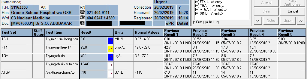
TSH: 42.9 mIU/L (0.51 – 4.30)

Free T4: 7.1 pmol/L (12.6 – 21.0)

Presenting Complaint

Patient presented with signs and symptoms of iron deficiency anemia.

History

The patient is a known hemophilia B carrier (Factor IX deficiency or Christmas disease) with menorrhagia and accompanying iron deficiency anemia.

Examination

The patient presented to the emergency rooms with symptoms of severe weakness and had occasional severe menorrhagia.

Unfortunately the physical examination details are not available.

Laboratory Investigations

  • Iron: 4.5 umol/L (9-30.4) Low
  • Transferrin 3.92 g/L (2-3.6) High
  • % Saturation 5% (15 -50) Low
  • Ferritin 8 ug/L (13- 68) Low

Other Investigations

Anti-Thyroglobulin (anti-TG) antibodies as well as Anti-Thyroid Peroxidase (anti-TPO) antibodies were positive in this patient.

These antibodies was suggested after the results of the TSH and reflexed free-T4 became available and hence after-requested.

Final Diagnosis

The pattern of significantly raised TSH with the significantly low free-T4 and the raised anti-TPO and anti-Thyroglobulin antibodies suggest primary hypothyroidism.

Take Home Message

It’s always worth doing a TSH to screen for thyroid disease when a patient presents with weakness / tiredness, irrespective of the age.

Primary hypothyroidism due to an auto-immune mediated destruction of the thyroid gland tissue is the most common hypothyroid condition and is confirmed by measuring the common anti-thyroid antibodies: Anti-TPO and Anti-TG antibodies. There is likely not much indication for performing these antibodies more than once after diagnosis of hypothyroidism and some clinicians argue it not necessary to even perform these antibody measurements.

Congenital hypothyroidism is one of the congenital disorders causing cretinism which is most preventable by newborn screening. Even though not likely congenital in this patient, it’s worth considering on the differential diagnosis.

The patient was on iron supplements as well as Factor IX injections. I was not aware of an association between Factor IX deficiency and Hypothyroidism, but my Haematology colleagues across the corridor told me the following:

Factor IX deficiency is an X-linked recessive disorder. This makes it unlikely for a female to get this disease. Auto-immune diseases is much more likely in females. There is also a form of Christmas disease where one produces antibodies to factor IX, which yields it inactive, hence presenting as Factor IX deficiency.

This, although unlikely, presents an interesting thought for this unusual presentation in this 16-year old female. Acquired deficiencies of most clotting factors have been described.

However, upon discussion with the attending clinician it seems that the patient did have a clear family history of Christmas disease, hence the presentation.


Elevated anti-Thyroglobulin Antibodies

HOSP # WARD Oudtshoorn Clinic
CONSULTANT George van der Watt & David Marais DOB/AGE 66y Male

Abnormal Result

Presenting Complaint

Mr. X, a 66 year old male, complained of chest pain, was seen at the Oudtshoorn Emergency department and a myocardial infarction was excluded by three serial point-of-care (POC) Troponin I results.

History

  • Known with hypothyroidism, but the cause was not defined yet.
  • On Eltroxin 150 ug daily PO
  • No other treatment.
  • Various stool analyses had been sent in for culture, with no definitive result.

Examination

Unfortunately not known.

Laboratory Investigations

Free T4: 24.6 pmol/L (7.6 – 16.1 pmol/L)

Anti-Thyroglobulin Antibody levels were elevated at 1944 U/mL (ref. <115 U/mL).

Other Investigations

Later, by retrospective viewing of the patient’s results it was revealed:

Total Cholesterol (TC) was elevated at 7.6 mmol/L. Hypothyroidism is associated with hypercholesterolemia. It can be concluded by the retrospective overview of results that upon an episode of hypothyroidism, the patient had hypercholesterolemia. This was most likely due to cessation of Thyroxine treatment, to whatever reason.

Index sample marked by the yellow shade. TC result which is raised (upper left corner) corresponds to the severely hypothyroid episode as revealed by the low T4 on that same sample.

Investigations also confirmatory for auto-immune hypothyroidism are:

  • Anti-Thyroid peroxidase antibodies
  • Anti-TSH receptor antibodies

Final Diagnosis

Auto-immune hypothyroidism

Take Home Messages

Interestingly, numerous patients with hypothyroidism is diagnosed at our Lipid Clinic at Groote Schuur Hospital. Patients are being referred for hypercholesterolaemia. Generally referral to this clinic happens when TC > 7.5 mmol/L. These patients are referred as presumed to have familial hypercholesterolaemia, but upon further work-up it is found that many of these patients have long-standing untreated hypothyroidism.

Prevalences of antithyroid antibodies as summarized by Up-to-date:

Image result for auto-immune hypothyroidism antibodies"


Thyroid Carcinoma

HOSP # WARD Nuclear Medicine
CONSULTANT   DOB/AGE 62 y Female

Abnormal Result

Thyroglobulin value of <0.1 ug/L upon signing results out.

Presenting Complaint

Patient presented with a neck “swelling” in October 2016. Systemic complaints were not inquired, but none was reported.

History

TFT: euthyroid since presentation when the thyroid functions were first tested in 2016.

Examination

A solitary thyroid nodule was found in the neck, moving with swallowing.

Laboratory Investigations

TFT: euthyroid upon presentation, which is clear from the table below.

  07/11/2019 08/08/2019 21/06/2019 20/02/2019 20/11/2018 26/09/2018 21/09/2018 05/07/2018 13/06/2018 29/05/2018 07/05/2018 08/01/2018 05/04/2017 26/10/2016
TSH δ- 1,49   δ- 7,07 H δ+62,97 H  0.01 L δ- 0.01 L        δ+88,52 H  3,84    6,35 H δ+ 9,88 H  2,86    4,05   δ+ 3,26    1,62  
Free T4  21,4   δ+ 22,3 H δ-  3.0 L δ- 29,8 H δ+ 42,7 H        δ-  3,1 L  17,3   CEGK  15,1                 δ+ 14,9      12  
Free T3                                                                                       4,8         
Thyroglobulin (3.5-77.0 ug/L)  <0.1 L  <0.1 L   0.1 L  <0.1 L δ- <0.1 L   7,9            8,8     7,6                                     
Anti-thyroglobulin Ab (<115 U/ml)   <10     <10     <10     <10     <10      11             14      11                                     

From results above it can be seen that the patient was euthyroid upon presentation (in 2016 and later until excision – highlighted in bold)

Hemithyroidectomy was done in May 2018, and another hemithyroidectomy in September 2018, thus the thyroglobulin after the first and second surgery was still detectable, but absent after September 2018.

Other Investigations

Ultrasound upon first presentation was indicative of a “suspicious nodule” in the thyroid gland.

Final Diagnosis

PATHOLOGICAL DIAGNOSIS – excision biopsy of nodule (17/01/2018)

Specimen A:
Right lobe of thyroid, hemithryoidectomy:
Encapsulated follicular variant of papillary thyroid carcinoma
Specimen B:
Right cervical lymph node (Level VI), biopsy:
Negative for malignancy (0/1)

PATHOLOGICAL DIAGNOSIS – left thyroid lobectomy (11/05/2018):

Left lobe of thyroid, excision: Negative for malignancy

PATHOLOGICAL DIAGNOSIS:

Right thyroid, lobectomy: Negative for malignancy (05/09/2018):

Take Home Messages

  • Thyroglobulin level is directly proportional to thyroid tissue present in vivo, hence is a useful tumour marker for thyroid cancer.
  • Anti-thyroglobulin antibody level is tested with Thyroglobulin to exclude false low values of thyroglobulin. If Anti-thyroglobulin Ab levels are increased with a decreased thyroglobulin level, the decrease in tumour marker is likely due to immunological clearance of the thyroglobulin and the result is hence likely false. This is the reason for the following canned text in TrakCare when the thyroglobulin level measures low:

The presence of thyroglobulin antibodies may interfere with the
thyroglobulin assay. The result of this test must be treated with reserve
if the patient has a positive thyroglobulin antibody test.

TrakCare canned text for low thyroglobulin

  • Upon total removal of the thyroid, thyroid replacement therapy is necessary, preferably slightly hyperthyroid levels to suppress TSH, and hence prevent tumour growth, as is also evidenced by this patient’s TSH and free T4 results.
  • As with most other tumour markers, thyroglobulin should be used for follow-up as a proxy of tumour size or regression.