Bilateral adrenal vein sampling

HOSP # Mr JB WARD Endocrine Department – CathLab – UCT private Hospital
CONSULTANT   Dr Jody Rusch DOB/AGE 53y Male

Abnormal Result

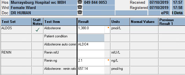
Upon authorizing blood results I came across a aldosterone result of 23300 pmol/L.

After a moment of brief anxiety, luckily I realized this was part of a series of tests performed by my colleagues in the Department.

Presenting Complaint

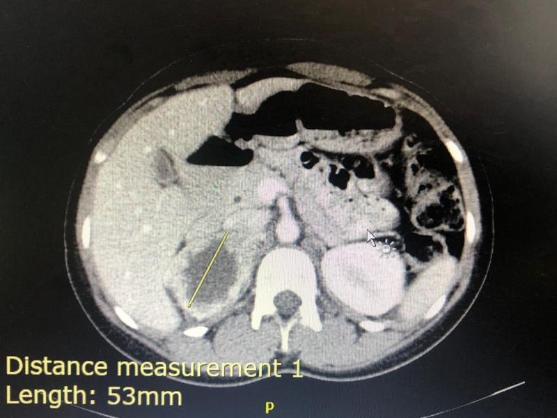
Medical complaint: Suspected Conn’s disease – right adrenal lesion/ irregular left adrenal gland.

History

The patient was confirmed to have primary hyperaldosteronism.

Unfortunately more information is not known. We were asked to assist with the sampling and the whole history weren’t available.

Examination

Not available

Laboratory Investigations

Table 1 – Results and calculations done in Excel.

Other Investigations

Not available for this patient.

Ideally one would need a CT with contrast beforehand to adequately visualize the positions of the adrenal veins, as this may aid in the canulation, especially of the right adrenal vein.

One needs to diagnose hyperaldosteronism (by an appropriate salt loading test) before proceeding to bilateral adrenal vein sampling.

Final Diagnosis

Interpretation

Definition Formula Clinical significance
Selectivity index PCC(side) / PCC (ivc) >cutoff confirms canulation of adrenal vein
>3 stimulated
>2 unstimulated
Lateralization index PAC/PCC (dom) : PAC/PCC (non-dom) >cutoff confirms laterilization of hyperaldo secretion
>4 stimulated
>2 unstimulated
Contralateral suppression index
(used if inadequate canulation)
PAC/PCC (non-dom) : PAC/PCC (ivc) <cutoff (<1 or <0.5 – sources differ) indicate ipsilateral
suppression
and suggest contralateral
aldosterone overproduction.
Table 1 – Interpretation of bilateral adrenal vein sampling.
PCC: plasma cortisol concentration, PAC: plasma aldosterone concentration, ivc: inferior vena cava or peripheral vein, dom: dominant side, non-dom: non-dominant side.

Selectivity index

Right: 1.0 (mean)

Left: 19.0 (mean)

These two results indicate that the left adrenal has been canulated adequately, but the right vein inadequately.

Lateralization index

Unable to comment because of the inadequate canulation of the right adrenal vein. If determined, it would very likely provide a false result.

Contralateral suppression index

1.5 : 1.8 = 0.8

This falls in between some of the referenced cutoffs (<1 and <0.5)

All of the other samples also fall somewhere in this range. Biochemically, these results suggests inadequate right sided venous sampling (a commonly described problem)

Take Home Message

  • Procedure is done in the Cath Lab
  • The patient received continuous synacthen infusion – as this improves the sensitivity (or perhaps rather specificity) of the test.
  • Done under imaging with contrast used for the localisation of the adrenal gland and adrenal vein
  • Sequential sampling technique used, generally > 20 mins infusion
  • Multi-disciplinary: nurses, anaesthetist, radiographer, intervention radiologists, students, chemical pathologists
  • Difficulty with sampling right side for both patients
  • Difficulty with interpreting results – most likely due to inadequate canulation of the right adrenal vein

Some important learning points

  1. Adrenal vein sampling may be a valuable tool that is underutilised
  2. Careful selection of patients essential – also patient should consent to surgical removal of the affected adrenal before this invasive procedure is initiated
  3. Inter-disciplinary approach is necessary
  4. Obtaining cosyntroponin (aka synacthen) can be difficult (Section 21), but recommended.
  5. Right adrenal access difficult: may require specific imaging. Recommended to start on the right or do simulataneous sampling.
  6. Adrenalectomy may be curative or help achieve better control of BP thus decrease associated morbidity and mortality in those with unilateral adenoma


Hyperaldosteronism with Hyperreninaemia in a 15 year old

HOSP # WARD Worcester Medical Ward
CONSULTANT  George van der Watt DOB/AGE 15 y Female

Abnormal Result

  • Aldosterone: 2320 pmol/L
  • Renin: 76.5 ng/L (Adult reference ranges – Supine: 1.7 – 23.9 ng/L; Upright: 2.7 – 27.7 ng/L)
  • Aldosterone:Renin Ratio : 30.3
  • The aldosterone:renin ratio (ARR) is a screening test for primary hyperaldosteronism and is most sensitive when both an absolute aldosterone > 350 pmol/L and an ARR > 118 pmol/ng is present.
  • Na: 138 mmol/L
  • K: 4.5 mmol/L
  • Urea 3.7 mmol/L
  • Creatinine: 49 umol/L
  • Total Calcium: 2.55 mmol/L

Urine dipstick 1+ protein

The urine protein:creatinine ratio was 0.044 g/mmol creat

Presenting Complaint

Patient presented with a 2 week history of blurry vision, intermittent headaches, hot flushes and mild intermittent epistaxis.

History

No known comorbidities

Multiple episodes of otitis media as a child

Presented with hypertension and evidence of target organ damage (retinopathy and left ventricular hypertrophy).

Examination

Vitals: HR 120, Resp. Rate 20, Temp 36.7 deg.C

BP (mmHg)

Right arm 144/92, Left arm 150/90

Right leg 176/74, Left leg 178/107

Gen: Not acutely / chronically ill, JACCOLD neg. Thyroid exam normal.

Funduscopy: Silver wiring, hard exudates, no haemorrhages, no papiloedema

CVS: bounding, peripherals pulses present. JVP raised, Undisplaced apex. Regular HR, no Radial/ femoral delays. Normal S1 & S2 with flowmurmur grade 2.

GIT: soft, non-tender. Ballotable left kidney, non-tender

Resp: Central Trachea, clear viscular breath sounds, no added sounds.

Neurological Exam: GCS 15/15, normal higher functions, no focal neurological signs.

BP control achieved with Doxazocin (increased to 4mg dly) and Atenolol (increased to 50mg dly)

Laboratory Investigations

TestItem Value Units Reference Range
Urine collection period 24 hours
Urine volume 1280 mL
Urine metanephrine 350 nmol/L
Urine normetanephrine 16350 nmol/L
dU metanephrine 448 nmol/24 hrs 167 – 938
dU normetanephrine 20928 (High) nmol/24 hrs 311 – 1562
Urine metanephrine : creat ratio 159 (High) nmol/mmol creat 17 – 88
Urine normetanephrine : creat ratio 7432 (High) nmol/mmol creat 23 – 176
Table 1 – Urine metanephrines (fractionated)

Other Investigations

ECG: Biatrial enlargement, left ventricular hypertrophy

Chest X-ray: Normal Cardio-thoracic index

Cardiac Ultrasound: Concentric left ventricular hypertrophy with preserved left ventricular ejection fraction. No valvular pathology.

KUB ultrasound: Similar kidney sizes. A mass with a cystic center was noted with no evidence of metastatic disease. Diagnosis suggested to be most likely a pheochromocytoma.

Final Diagnosis

Right-sided phaeochromocytoma

Take Home Message

Aldosterone : Renin ratio cannot be looked at alone. A raised value in either of the Aldosterone and Renin should be investigated further, especially if severely deranged like in this case.

Always investigate hypertension in a child until the cause is found. Hypertension in a child is not normal.

3 consecutive 24 hour urine collection samples are recommended for diagnosis of phaeochromocytoma as some tumours only secrete epinephrines / norepinephrines episodically. In this case however it was not necessary as the case was clear with a markedly raised dU-normetanephrine level.

Screening for pheochromocytoma is an essential part of the workup for secondary hypertension. Urinary vanillyl mandelic acid (VMA) was traditionally used to diagnose phaeochromocytoma. It has a low sensitivity (60-70%). Later, catecholamines measurement in plasma (PCAT) and urine (UCAT) emerged as useful tests. The sensitivity of catecholamines is limited by their episodic release from the tumour cells. The sensitivity ranges from 76-86 % for PCAT and UCAT and the specificity is around 81-99 %.

Metanephrines are methylated metabolites of catecholamines.

Metanephrines are secreted continuously from the tumour cells, independent of the intermittent release of catecholamines. The metanephrines are metabolized by conjugation, primarily in the hepatomesentric organs. Plasma metanephrines (pMN) are measured in the free form (not routinely offered in South Africa) whereas urinary metanephrines (uMN) represent mainly the conjugated form. Hence compared to pMN, uMN is less specific. Studies have shown that plasma free metanephrines have a sensitivity of 96-100 per cent and specificity of 85-100 % superior to that of uMN which has a sensitivity of 93-99.6 per cent and specificity of 71-77 per cent.

Previous methods used colorimetry or spectrophotometry as total MET (metanephrine + normetanephrines) which includes a combined measurement of metanephrine (MN) and normetanephrine (NMN). These methods were superseded by liquid chromatographic assays (LC) that allow individual measurement of MN and NMN.

At Red Cross Hospital Laboratory we use a gas chromatography with mass spectrometry, which is not so widely used for fractionated metanephrines. An isotope dilution method is employed, bringing the method up to internationally recognized standards and the quality assurance of the method at the Red Cross Chemistry lab performs well on the international EQA scheme used.


Hyperaldosteronism

HOSP # WARD Murraysburg Hospital, Female Ward
CONSULTANT   DOB/AGE 51 y female

Abnormal Result

Aldosterone: 1380 pmol/L

Renin: 2.1 ng/L

Aldosterone: Renin ratio: 657.14 pmol/ng

Presenting Complaint

Uncontrolled Hypertension, unresolved on maximum dose of 3 antihypertensives.

History

Examination

Laboratory Investigations

Other Investigations

Urine electrolytes

Serum Results

Date Sodium mmol/L Potassium mmol/L eGFR ml/min GGT U/L Chol mmol/L TSH mIU/L T4 pmol/L FreeT3 pmol/L Cort nmol/L
21/04/2015          2,8   >60           5,07                              
30/11/2015          3,8     >60           4,53                              
15/11/2016          >60           4,04                              
20/03/2017                 >60           4,36                              
05/06/2018   144  3,4    56           4,39    1,79    11,9       5     394  
20/08/2018   131 4,6    42                                            
21/08/2018                                                               
24/08/2018                                                               
26/08/2018                                                               
26/08/2018                                                               
26/09/2019   139   2,4    45                  0.81                       
27/09/2019   142   2,6    43                                            
01/10/2019                                                               
02/10/2019   139   2,9    40                 CEGK                
03/10/2019                                                               
07/10/2019   138   3,9      38                                            
31/10/2019   139   1,9    30      28                                     

Urine metanephrines

Urine collection period 24  h       Reference value
Urine volume 3080 ml       
Ucreat   2,2 mmol/L       
Umetadren   160 nmol/L       
Unormetadren   870  nmol/L
dUmetadren   493  nmol/24h 152-913
dUnormetadren  2680 nmol/24h 699-2643
Umetadren:cr    73  nmol/mmol creat 17-91
Unormetad:cr   395 nmol/mmol creat 75-309

Final Diagnosis

Primary hyperaldosteronism causing secondary hypertension with accompanying renal injury.

Take Home Messages

Reference Ranges for Aldosterone:

  • Upright 70 – 1066 pmol/L
  • Supine 49 – 643 pmol/L

Screening for primary hyperaldosteronism: most sensitive when >350 pmol/L

Reference Ranges for Renin:

  • Upright: 2.7 – 27.7 ng/L
  • Supine: 1.7 – 23.9 ng/L

Beta-blockers suppress renin levels and should be stopped 2 weeks before testing.

Aldosterone: Renin Ratio:

Most sensitive when the ratio is >118 pmol/ng.

Effects of hyperaldosteronism

  • One’s expectation is a high serum sodium, but since it normalizes with an increase in fluid volume, hence hypertension as in this case, there is normal sodium.
  • Low serum potassium due to loss in urine, although this can also be normal.
  • Increased urine potassium concentration (>30 mmol/L) in a random urine specimen suggests increased mineralocorticoid effect.
  • The renin:aldosterone ratio is used to compensate for the increase in aldosterone which is caused by an increase in renin (for instance which is caused by hypovolemia or low blood pressure).
  • Some studies recently published are suggesting that the prevalence of hyperaldosteronism are significantly more than was (and is) thought, and hence urinary (24 hour) aldosterone measurement may be more accurate to screen for hyperaldosteronism. The authors of recent estimates of the prevalence of hyperaldosteronism are of opinion that hyperaldosteronism may be the cause of around 10% of unexplained “essential” hypertensives (see attached articles).