Hyperaldosteronism with Hyperreninaemia in a 15 year old
| HOSP # | WARD | Worcester Medical Ward | |
| CONSULTANT | George van der Watt | DOB/AGE | 15 y Female |
Abnormal Result
- Aldosterone: 2320 pmol/L
- Renin: 76.5 ng/L (Adult reference ranges – Supine: 1.7 – 23.9 ng/L; Upright: 2.7 – 27.7 ng/L)
- Aldosterone:Renin Ratio : 30.3
- The aldosterone:renin ratio (ARR) is a screening test for primary hyperaldosteronism and is most sensitive when both an absolute aldosterone > 350 pmol/L and an ARR > 118 pmol/ng is present.
- Na: 138 mmol/L
- K: 4.5 mmol/L
- Urea 3.7 mmol/L
- Creatinine: 49 umol/L
- Total Calcium: 2.55 mmol/L
Urine dipstick 1+ protein
The urine protein:creatinine ratio was 0.044 g/mmol creat
Presenting Complaint
Patient presented with a 2 week history of blurry vision, intermittent headaches, hot flushes and mild intermittent epistaxis.
History
No known comorbidities
Multiple episodes of otitis media as a child
Presented with hypertension and evidence of target organ damage (retinopathy and left ventricular hypertrophy).
Examination
Vitals: HR 120, Resp. Rate 20, Temp 36.7 deg.C
BP (mmHg)
Right arm 144/92, Left arm 150/90
Right leg 176/74, Left leg 178/107
Gen: Not acutely / chronically ill, JACCOLD neg. Thyroid exam normal.
Funduscopy: Silver wiring, hard exudates, no haemorrhages, no papiloedema
CVS: bounding, peripherals pulses present. JVP raised, Undisplaced apex. Regular HR, no Radial/ femoral delays. Normal S1 & S2 with flowmurmur grade 2.
GIT: soft, non-tender. Ballotable left kidney, non-tender
Resp: Central Trachea, clear viscular breath sounds, no added sounds.
Neurological Exam: GCS 15/15, normal higher functions, no focal neurological signs.
BP control achieved with Doxazocin (increased to 4mg dly) and Atenolol (increased to 50mg dly)
Laboratory Investigations
| TestItem | Value | Units | Reference Range |
|---|---|---|---|
| Urine collection period | 24 | hours | |
| Urine volume | 1280 | mL | |
| Urine metanephrine | 350 | nmol/L | |
| Urine normetanephrine | 16350 | nmol/L | |
| dU metanephrine | 448 | nmol/24 hrs | 167 – 938 |
| dU normetanephrine | 20928 (High) | nmol/24 hrs | 311 – 1562 |
| Urine metanephrine : creat ratio | 159 (High) | nmol/mmol creat | 17 – 88 |
| Urine normetanephrine : creat ratio | 7432 (High) | nmol/mmol creat | 23 – 176 |
Other Investigations
ECG: Biatrial enlargement, left ventricular hypertrophy
Chest X-ray: Normal Cardio-thoracic index
Cardiac Ultrasound: Concentric left ventricular hypertrophy with preserved left ventricular ejection fraction. No valvular pathology.
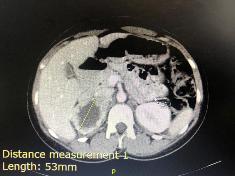
KUB ultrasound: Similar kidney sizes. A mass with a cystic center was noted with no evidence of metastatic disease. Diagnosis suggested to be most likely a pheochromocytoma.
Final Diagnosis
Right-sided phaeochromocytoma
Take Home Message
Aldosterone : Renin ratio cannot be looked at alone. A raised value in either of the Aldosterone and Renin should be investigated further, especially if severely deranged like in this case.
Always investigate hypertension in a child until the cause is found. Hypertension in a child is not normal.
3 consecutive 24 hour urine collection samples are recommended for diagnosis of phaeochromocytoma as some tumours only secrete epinephrines / norepinephrines episodically. In this case however it was not necessary as the case was clear with a markedly raised dU-normetanephrine level.
Screening for pheochromocytoma is an essential part of the workup for secondary hypertension. Urinary vanillyl mandelic acid (VMA) was traditionally used to diagnose phaeochromocytoma. It has a low sensitivity (60-70%). Later, catecholamines measurement in plasma (PCAT) and urine (UCAT) emerged as useful tests. The sensitivity of catecholamines is limited by their episodic release from the tumour cells. The sensitivity ranges from 76-86 % for PCAT and UCAT and the specificity is around 81-99 %.
Metanephrines are methylated metabolites of catecholamines.
Metanephrines are secreted continuously from the tumour cells, independent of the intermittent release of catecholamines. The metanephrines are metabolized by conjugation, primarily in the hepatomesentric organs. Plasma metanephrines (pMN) are measured in the free form (not routinely offered in South Africa) whereas urinary metanephrines (uMN) represent mainly the conjugated form. Hence compared to pMN, uMN is less specific. Studies have shown that plasma free metanephrines have a sensitivity of 96-100 per cent and specificity of 85-100 % superior to that of uMN which has a sensitivity of 93-99.6 per cent and specificity of 71-77 per cent.
Previous methods used colorimetry or spectrophotometry as total MET (metanephrine + normetanephrines) which includes a combined measurement of metanephrine (MN) and normetanephrine (NMN). These methods were superseded by liquid chromatographic assays (LC) that allow individual measurement of MN and NMN.
At Red Cross Hospital Laboratory we use a gas chromatography with mass spectrometry, which is not so widely used for fractionated metanephrines. An isotope dilution method is employed, bringing the method up to internationally recognized standards and the quality assurance of the method at the Red Cross Chemistry lab performs well on the international EQA scheme used.