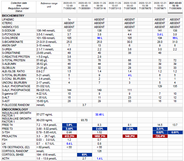
Hyperprolactinemia >1000

HOSP # WARD Neurosurgery
CONSULTANT Dr. Jody Rusch   DOB/AGE 10 year female

Abnormal Result

Prolactin >1000 ug/L

Presenting Complaint

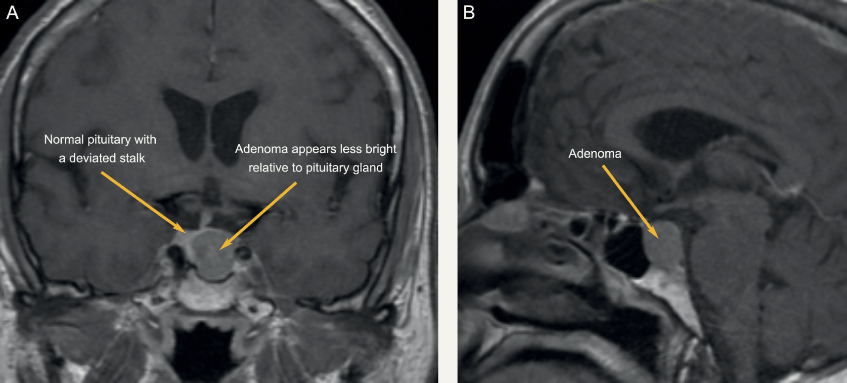
Patient presented at 7 years of age with galactorrhea and visual field defects.

History

Patient had a craniotomy for debulking of the adenoma. This was opposed to the usual transsphenoidal more non-invasive route of pituitary adenoma surgery. She was initiated on Cabergoline 1 g twice weekly for suppression of the tumour size.

It was also noted during surgery that the tumour was extremely vascular with much bleeding and the neurosurgeons struggled to mobilize it to adequately get it separated from the optic chiasm. Some portion of the tumour was left in situ during surgery as this was too big a risk for trying to excise.

A biopsy was also taken.

Examination

Patient subsequently developed severe intracranial edema after surgery in the ICU.

Laboratory Investigations

Other Investigations

Histology

Frozen section – pituitary adenoma. GROSS DESCRIPTION: Specimen labelled tumour. Specimen consists of 2 fragments of tissue, larger measuring 4x3mm. HISTOLOGY: Sections show tumour tissue composed of nests of monotonous cells with intervening fibrous septae. The cells have round nuclei and abundant eosinophilic cytoplasm. The nuclei have stippled chromatin with inconspicuous nucleoli. No mitotic activity or necrosis is seen. Immunohistochemistry: Synaptophysin:Positive Prolactin: Positive LH: Negative FSH: Negative GH: Negative TSH: Negative ACTH: Negative CONCLUSION: Pituitary, mass, excision: – Pituitary adenoma with an immunohistochemical profile compatible with a prolactinoma.

Final Diagnosis

Pituitary Macroadenoma

Take Home Message

Cabergoline, sold under the brand name Dostinex among others, is a dopaminergic medication used in the treatment of high prolactin levels, prolactinomas, Parkinson’s disease, and for other indications. It is taken by mouth. Cabergoline is an ergot derivative and a potent dopamine D₂ receptor agonist.

Lactotroph adenomas (prolactinomas) are more amenable to pharmacologic treatment than any other kind of pituitary adenoma because of the availability of dopamine agonists, which usually decrease both the secretion and size of these tumors. For the minority of lactotroph adenomas that do not respond to dopamine agonists, other treatments must be used. Hyperprolactinemia due to nonadenoma causes should also be treated if it causes hypogonadism.

There are two principal reasons why patients with hyperprolactinemia may need to be treated: existing or impending neurologic symptoms due to the large size of a lactotroph adenoma, and hypogonadism or other symptoms due to hyperprolactinemia, such as galactorrhea.

A third indication is in women with mild hyperprolactinemia and normal cycles who are trying to conceive as they may have subtle luteal phase dysfunction.


A case of amenorrhoea in a 17-year old female

HOSP # MRN94883340 WARD Paeds Endocrine Clinic
CONSULTANT   Jody Rusch / Ariane Spitaels DOB/AGE 17 year female

Abnormal Result

Prolactin 51.1 ug/L

Monomeric Prolactin 36.2 ug/L

Presenting Complaint

Amenorrhoea (more details unknown)

History

The patient presented with a tempoparietal tumour and had received two episodes of radiotherapy – was asked by the oncologists to be reviewed by the Endocrinologists.

Mother stopped epilim (reason unknown)

Patient currently has amenorrhoea (unknown whether it is primary or secondary)

Examination

Residual right hemiplegia

Unfortunately no other facts about the physical examination are known

Laboratory Investigations

  • Normal TFT:
    • TSH 1.7 mIU/L (0.51 – 4.3)
    • Free T4 16.2 (12.6 – 21.0)
  • Cort 11am 330 nmol/L
  • FSH 3.8 IU/L
  • LH 2.4 IU/L
  • E3 106 pmol/L
  • Prol 51.1 ug/L
  • Monomeric Prolactin 36.2 ug/L
  • Recovery: 70.8%

Other Investigations

Proposed investigations:

  • Pregnancy test (most common cause of amenorrhoea)
  • Ovarian ultrasound to exclude early-onset PCOS (which may become a diagnosis of exclusion)
  • History about prior amenorrhoea
  • Brain MRI to visualize pathology in the cranium

Final Diagnosis

Hyperprolactinemia – likely causing amenorrhoea – cause yet to be determined

Take Home Message

Hyperprolactinemia is perhaps one of the most common problems in clinical endocrinology. It relates with various aetiologies (see below), the clarification of which requires careful history taking and clinical assessment. Analytical issues (presence of macroprolactin or of the hook effect) need to be taken into account when interpreting the prolactin values. Medications and sellar/parasellar masses (prolactin secreting or acting through “stalk effect”) are the most common causes of pathological hyperprolactinaemia. Hypogonadism and galactorrhoea are well-recognized manifestations of prolactin excess, although its implications on bone health, metabolism and immune system are also expanding. Treatment mainly aims at restoration and maintenance of normal gonadal function/fertility, and prevention of osteoporosis; further specific management strategies depend on the underlying cause.

The main physiological causes of hyperprolactinemia:

  • Ovulation
  • Pregnancy
  • Breastfeeding
  • Stress
  • Exercise
  • Nipple stimulation or chest wall injury

Pathological

  • Prolactin-secreting pituitary adenoma
  • “Stalk-effect” from sellar / parasellar lesions
  • Renal failure
  • Liver cirrhosis
  • Primary hypothyroidism
  • Polycystic Ovarian Syndrome
  • Seizures

Pharmacological

  • Antipsychotics / neuroleptics
  • Antidepressants
  • Antiemetics
  • Opioids
  • Antihypertensives

It is clear in this case that the history is quite important in any patient in whom hyperprolactinemia is detected, since a vast array of causes exist.

For an excellent review on prolactin: https://www.ncbi.nlm.nih.gov/pmc/articles/PMC6947286/

For another case of high prolactin see:


Rapidly decreasing Prolactin result

HOSP # WARD Endocrinology Clinic
CONSULTANT John Stanfliet   DOB/AGE 36 y Female

Abnormal Result

A low prolactin result was obtained in a patient in whom a macroadenoma was suspected:

Prolactin: 1.3 mIU/L

Presenting Complaint

The patient presented with headache and decreased visual acuity (more specifically peripherally).

History

There were bilateral galactorrhoea, amenorrhoea, and as noted above, headache and visual disturbances.

The patient had received Cabergoline (a dopamine receptor agonist on D2 receptors) for the past 4 months.

Examination

As above

Laboratory Investigations

Date Prolactin (mIU/L)
02/2019 106 (Recovery of 80% following PEG precipitation)
05/2019 135
06/2019 85
08/2019 1.3 (1.59 with a 1:10 dilution; 3.94 with a 1:50 dilution)
Prolactin Results

Other Investigations

MRI Head was booked for the following week. Interestingly, even in prolactin secreting tumours, the correlation between tumour size and prolactin level is limited. MRI head remains a vital investigation.

Final Diagnosis

An Introduction to Sellar Masses (Chapter 10) - Clinical Neuroendocrinology
Pituitary Macroadenoma

Take Home Message

During pregnancy the concentration of prolactin rises under the influence of elevated estrogen and progesterone production. The stimulating action of prolactin on the mammary gland leads post partum to lactation. Hyperprolactinemia (in men and women) is the main cause of fertility disorders. The determination of prolactin is utilized in the diagnosis of anovular cycles, hyperprolactinemic amenorrhea and galactorrhea, gynecomastia and azoo-spermia. Prolactin is also determined when breast cancer and pituitary tumors are suspected. As in this case, a pituitary tumour was suspected, hence the repeated prolactin results.

As was noted in another short case, our assay on the Roche platform does measure all forms of prolactin, and when a high result is obtained (above the gender-specific reference range) it is recommended to measure the recovery after PEG precipitation.

Figure 1

Dr. John Stanfliet (pathologist at Pathcare) repied to the above case with very valuable comments:

  • We use Beckman Coulter DxI, an immunoassay that is not affected by macroprolactin (I’ve include an article that shows this).
  • Even in prolactin secreting tumours, the correlation between tumour size and prolactin level is limited.  MRI head remains a vital investigation.
  • Some prolactin secreting tumours also secrete other pituitary hormones such as growth hormone.
  • I would ascribe the reduction in PRL to the Carbegoline and wonder whether the dose has been increased.
  • Dr. Pete Berman would often suggest a mixing study: find a sample with high PRL, mix it 50/50 with this sample, and measure it to see whether there is some interferant in this sample. 


Prolactin

HOSP # WARD ENT Clinic
CONSULTANT   DOB/AGE 35 Y Male

Abnormal Result

Prolactin 10 986.0 ug/L (4-15.2) 

Dilutions:

1/10  >4700;

1/100 = 10821;

1/50 = 10 986.

Presenting Complaint

Epistaxis

History

Patient with epistaxis referred to the ENT specialist clinic.  No relevant medication history.

Examination

35 y male with a large left post-nasal space mass, a vascular mass involving the pituitary fossa.

?NBL (non-benign lesion)

?Sinonasal malignancy

?Pituitary Tumour

Laboratory Investigations

TSH 0.91 pmol/L (0.27-4.20)

Free T4 15.7 pmol/L (12-22)

FSH 0.8 IU/L ↓ (1.5-12.4)

LH 0.2 IU/L ↓ (1.7-8.6)

Testosterone 0.2 nmol/L ↓ (8.6-29.0)

PTH 1.7 pmol/L (1.6-6.9)

Prolactin measuring method:

The Elecsys prolactin sandwich immunoassay uses two monoclonal
antibodies directed against human prolactin.

R1 = biotinylated antibody – recognizes the N-terminal end of the
prolactin molecule

R2 – ruthenium complexed antibody probably reacts with a region in the
middle of the prolactin molecule.

1st incubation: a biotinylated monoclonal prolactin-specific
antibody and a monoclonal prolactin-specific antibody labeled with a ruthenium
complex form a sandwich complex.

2nd incubation: after addition of streptavidin-coated
microparticles, the complex becomes bound to the solid phase via interaction of
biotin and streptavidin.

Reaction mixture aspirated into the measuring cell where microparticles
are magnetically captured into the surface of the electrode.   Unbound substances are then removed with
ProCell. 

Application of a voltage to the electrode then induces
chemiluminescent emission which is measured by a photomultiplier, results
calculated by a standard curve.

Other Investigations

Monomeric prolactin – 7744 ug/L (70% recovery after PEG precipitation)

Biopsy: confirmed tumour stained strongly positive
with prolactin suggesting a prolactinoma.

Final Diagnosis

Pituitary Macroprolactinoma

Take Home Messages

Sandwich immunoassays are prone to high dose hook-effect. There are
various ways to overcome this effect. (This will later be expanded on – see AFP
/ Beta-HCG).

Prolactin appears in the serum as:

  1. Active monomeric
    prolactin (“little”) (80%) 23kDa
  2. Inactive dimeric
    prolactin (“big”) (5-20%) 50-60kDa
  3. Low activity
    tetrameric prolactin (“big-big”) (0.5-5%) 150-170kDa 

Precipitation by PEG yields the active monomeric
prolactin, expressed as a percentage recovery after precipitation.  Big-big prolactin consists of an
antigen-antibody complex of monomeric prolactin-immunoglobulin G and is defined
as macroprolactin.  This has a long
half-life in blood when compared to normal prolactin and gives false high
readings of prolactin, leading to unnecessary investigations in certain
cases.  A high prolactin should thus be
confirmed by doing a PEG precipitation.