Possible Heterophile antibodies

HOSP # MRN77113313 WARD Endocrinology OPD
CONSULTANT   Dr. Jody Rusch / Dr. Khalid Aligail DOB/AGE 21y Female

Abnormal Result

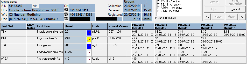
The TSH stayed elevated on our assay (Roche Cobas 6000) with a high-normal free T4.

Presenting Complaint

The patient was seen at the endocrinology OPD for follow-up of her thyroid function tests and review of medications. No acute complaints were noted, but some interesting thyroid function results became known.

History

Previous multinodular goiter with thyrotoxicosis.  Had a complete thyroidectomy March-May 2020.

History of asthma, exema and “other allergies”. 

Current dose of eltroxin = 1.6 ug/kg ~ 100ug/day PO.  The patient (and doctor) declares good compliance to Rx.

Examination

Patient did not have any signs or symptoms of hypo or hyperthyroidism according to the endocrinologist.

Laboratory Investigations

Date 03/05/2021 26/04/2021 23/02/2021 23/10/2020 27/08/2020 08/05/2020 09/03/2020
TSH (uIU/mL) (0.27 – 4.2) •15,17 H 18,54 H 13.10 H 21,61 H 32,19 H δ+>100.00 H  7,72 H
Free T4 (pM) (12.0-22.0)  17,8    17,7    18,3   δ+ 16.0    11,8 L δ-  9,7 L δ+ 13,8  
Free T3 (pM) (3.1 – 6.8)   4,3            4,2                              
Table 1 – Thyroid function tests, cumulative history – Results as on Roche Cobas 6000

The TSH seems to have stayed elevated on our assay (Roche Cobas 6000) with a high-normal free T4.  The free T3 is normal (which I advised should be measured to assess conversion between the hormones). I also sent the sample to Green Point Laboratory where a Beckman DXi analyser is used with a different antibody set of reagents and a different reference range.

Date 03/05/2021
TSH (uIU/mL) 15.4 (0.38-5.33)
Free T4 (pM) 13.6 (7.86-14.41)
Free T3 (pM) 4.3 (3.8-6.0)
Table 2 – results of sample on 03/05/2021 ran on the Beckman DXi

Other Investigations

Auto-immune markers have been requested, since the patient was having prolonged iron deficiency, becoming anaemic, and the clincian raised a suspician of likely celiac disease.

Final Diagnosis

The diagnosis is still unsure, but the likely differential diagnosis is:

  1. Decrease in deiodinase activity due to some reason – there are many causes.
  2. Decrease in absorbtion of Levothyroxine

Take Home Message

Interference in thyroid function tests are commonly enquired about, especially by endocrinologists. This represents a big portion of our non-routine work and often quite a portion of time is spent on education of clinicians who do not necessarily have a laboratory background. Often, esoteric tests are requested which may not be warranted by the clinical scenario.

This case demonstrates a query raised by a clinician about possible interference in the TFT’s which is warranted. It is important to rule out as best one can, interference in the laboratory assay.

Various ways of determining whether interference is the culprit are:

  • Dilution of the sample (in assays where the sample may be diluted – unsuitable for free-T4 as dilution will affect the “free” portion of hormone)
  • Running the test on another methodology
  • Running the test on another analyzer of the same methodology, but with slight differences, such as a different manufacturer of detection antibodies (e.g. Roche vs. Abbott vs. Siemens vs. Beckman vs. Ortho)
  • Precipitating the antibodies e.g. desalting, or PEG-precipitation.
  • Binding the antibodies, e.g. protein G or Protein A
  • Using of “blocking tubes” which is a proprietary blood collection tube to bind antibodies


Thyroid Carcinoma

HOSP # WARD Nuclear Medicine
CONSULTANT   DOB/AGE 62 y Female

Abnormal Result

Thyroglobulin value of <0.1 ug/L upon signing results out.

Presenting Complaint

Patient presented with a neck “swelling” in October 2016. Systemic complaints were not inquired, but none was reported.

History

TFT: euthyroid since presentation when the thyroid functions were first tested in 2016.

Examination

A solitary thyroid nodule was found in the neck, moving with swallowing.

Laboratory Investigations

TFT: euthyroid upon presentation, which is clear from the table below.

  07/11/2019 08/08/2019 21/06/2019 20/02/2019 20/11/2018 26/09/2018 21/09/2018 05/07/2018 13/06/2018 29/05/2018 07/05/2018 08/01/2018 05/04/2017 26/10/2016
TSH δ- 1,49   δ- 7,07 H δ+62,97 H  0.01 L δ- 0.01 L        δ+88,52 H  3,84    6,35 H δ+ 9,88 H  2,86    4,05   δ+ 3,26    1,62  
Free T4  21,4   δ+ 22,3 H δ-  3.0 L δ- 29,8 H δ+ 42,7 H        δ-  3,1 L  17,3   CEGK  15,1                 δ+ 14,9      12  
Free T3                                                                                       4,8         
Thyroglobulin (3.5-77.0 ug/L)  <0.1 L  <0.1 L   0.1 L  <0.1 L δ- <0.1 L   7,9            8,8     7,6                                     
Anti-thyroglobulin Ab (<115 U/ml)   <10     <10     <10     <10     <10      11             14      11                                     

From results above it can be seen that the patient was euthyroid upon presentation (in 2016 and later until excision – highlighted in bold)

Hemithyroidectomy was done in May 2018, and another hemithyroidectomy in September 2018, thus the thyroglobulin after the first and second surgery was still detectable, but absent after September 2018.

Other Investigations

Ultrasound upon first presentation was indicative of a “suspicious nodule” in the thyroid gland.

Final Diagnosis

PATHOLOGICAL DIAGNOSIS – excision biopsy of nodule (17/01/2018)

Specimen A:
Right lobe of thyroid, hemithryoidectomy:
Encapsulated follicular variant of papillary thyroid carcinoma
Specimen B:
Right cervical lymph node (Level VI), biopsy:
Negative for malignancy (0/1)

PATHOLOGICAL DIAGNOSIS – left thyroid lobectomy (11/05/2018):

Left lobe of thyroid, excision: Negative for malignancy

PATHOLOGICAL DIAGNOSIS:

Right thyroid, lobectomy: Negative for malignancy (05/09/2018):

Take Home Messages

  • Thyroglobulin level is directly proportional to thyroid tissue present in vivo, hence is a useful tumour marker for thyroid cancer.
  • Anti-thyroglobulin antibody level is tested with Thyroglobulin to exclude false low values of thyroglobulin. If Anti-thyroglobulin Ab levels are increased with a decreased thyroglobulin level, the decrease in tumour marker is likely due to immunological clearance of the thyroglobulin and the result is hence likely false. This is the reason for the following canned text in TrakCare when the thyroglobulin level measures low:

The presence of thyroglobulin antibodies may interfere with the
thyroglobulin assay. The result of this test must be treated with reserve
if the patient has a positive thyroglobulin antibody test.

TrakCare canned text for low thyroglobulin

  • Upon total removal of the thyroid, thyroid replacement therapy is necessary, preferably slightly hyperthyroid levels to suppress TSH, and hence prevent tumour growth, as is also evidenced by this patient’s TSH and free T4 results.
  • As with most other tumour markers, thyroglobulin should be used for follow-up as a proxy of tumour size or regression.