A possible case of glycerokinase deficiency

HOSP # WARD Ward B2
CONSULTANT   George van der Watt DOB/AGE 3 month

Abnormal Result

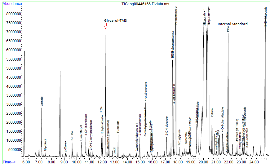
Glycerol which is significantly raised on urine organic acid analysis.

Figure 1 – Chromatogram. The high levels of Glycerol (with TMS – trimethyl silyl derivative) which is >0.5 the peak height of the internal standard (PCA – pentadecanoic acid).
Figure 2 – Mass spectrum of the peak as indicated by Glycerol TMS above in Figure 1.
Figure 3 – Follow up gas chromatogram without KY-jelly
Figure 4 – Mass spectrum in the peak marked as “Glycerol-TMS” from figure 3.

Presenting Complaint

Patient is a 3 month old male with signs and symptoms of sepsis.

History

Patient presented with significant failure to thrive.

Laboratory Investigations

Triglycerides : 4.47 mmol/L

Other Investigations

Faecal elastase 81 ug/g stool

Reference range (adults and children > 1 month):

  • > 200 ug elastase/g stool: Normal exocrine pancreatic function
  • 100-200 ug elastase/g stool: Moderate/mild pancreatic insufficiency
  • < 100 ug elastase/g stool: Severe exocrine pancreatic insufficiency

These ranges apply to formed stool samples. Watery stool samples may yield spuriously low elastase results due to dilution, and a formed stool sample should be sent for re-analysis.

Final Diagnosis

Glycerol contamination of the skin – as excluded by the repeat analysis.

Take Home Message

  • Glycerol (glycerine) is a common contaminant of urine organic acids due to being present in various skin products / creams. Contamination can be eliminated by thorough cleaning of the perineum with normal saline or doing an “in-out” catheterization procedure for urine collection in neonates. Interestingly glycerol is also one of the main ingredients in KY jelly, a common lubricant use for catheterization.
  • High glycerol in serum will present with a falsely high triglyceride level on most routine chemistry analysers due to the inherent enzymatic conversion of triglycerides to glycerol before further steps to measurement.
Glycerol Kinase - an overview | ScienceDirect Topics
Figure 3 – Explanation of triglyceride determination by amperometric detection.
  • Sepsis is more common than inherited metabolic diseases and so is pre-analytical caveats such as glycerol contamination of the perineal skin.


Hypernatremia

HOSP # WARD Red Cross Children’s Hospital ICU
CONSULTANT Dr. S
Prof. G
DOB/AGE 14 day old Neonate

Abnormal Result

Sodium = 198 mmol/L (H) (136-145)

Presenting Complaint

1 day of poor feeding.  Child passing very hard/ dark brown stool for the preceding 10 days.

History

Birth weight @ term: 3.380kg.  Delivered vaginally after induction of labour because of spontaneous rupture of membranes at 40 weeks gestation. Discharged home without any problems after 1 day.

Examination

On arrival at district hospital: Temp: 38oC, Sats 96% on Nasal O2, Finger prick glucose: 10mmol/L, Capillary refill time: 6 seconds,

HR: 140bpm. 

Blood gas:

pH: 7.26,

BE -16.3,

pCO­2 3.2 kPa,

Na 190.

Weight: 2.2kg (birth weight: 3.380 kg, thus 35% weight loss)

Laboratory Investigations

Other Investigations

Urine organic acid analysis by GCMS demonstrates elevation of the liver markers 4-OH-phenyllactate and 4-OH-phenylpyruvate together with lactaturia. Succinylacetone, a marker for tyrosinaemia type 1 is absent. Moderate ketonuria with elevated dicarboxylic acids C6, C8, and C10 is also present, these changes suggest a lipolytic response to catabolic or fasting stress or hypoglycaemia together with underlying hepatic dysfunction with lactataemia but are non-specific for an IMD per se. 

Final Diagnosis

Patient was pure water depleted with a sodium concentration of 198 mmol/L.  The mother was not lactating adequately despite the infant sucking well, evidenced by the fact that when expressed breast milk was tried, there was too little milk for the baby to drink.  The nurses’ notes confirmed this finding.  This finding also confirms the failure to produce stool volume and the normal urine organic acid profile with evidence of starvation / fasting stress.

Take Home Messages

When considering a patient with high plasma sodium concentration it is
important to bear in mind:

  1. Hypernatremia does not necessarily indicate an excess of extracellular sodium.  Except in rare cases of salt overload most patient with hypernatremia have a deficiency of both water and sodium, with the water deficiency being proportionally higher than that of sodium.
  2. Patients become hypernatremic because the water lost from the body exceed the intake and there is negative fluid balance.  The amount of water which a person can drink generally exceeds by far the amount lost from the body in most pathological fluid-losing disorders, eg. Diarrhoea, sweating.  Patients thus become hypernatremia due to:
    1. Too old, young or sick to drink
    2. Obstruction of oesophagus
    3. Disorders of thirst centre
    4. No access to water                          

Ref: Walmsley – Cases in Chemical Pathology 4th
ed.

It is also important:

  1. To calculate the Osmolar gap( difference between calculated and measured osmolarity)
  2. U:P osmol (>1 = hypotonic fluid depletion, pure water loss or salt gain; ~1 = osmotic diuresis; <1 = diabetes insipidus ~the various causes of nephrogenic and neurogenic DI)