A case of persistent hypocalcemia

HOSP # MRN63985901 WARD Medical Ward
CONSULTANT Dr. Heleen Vreede   DOB/AGE 51 year Female

Abnormal Result

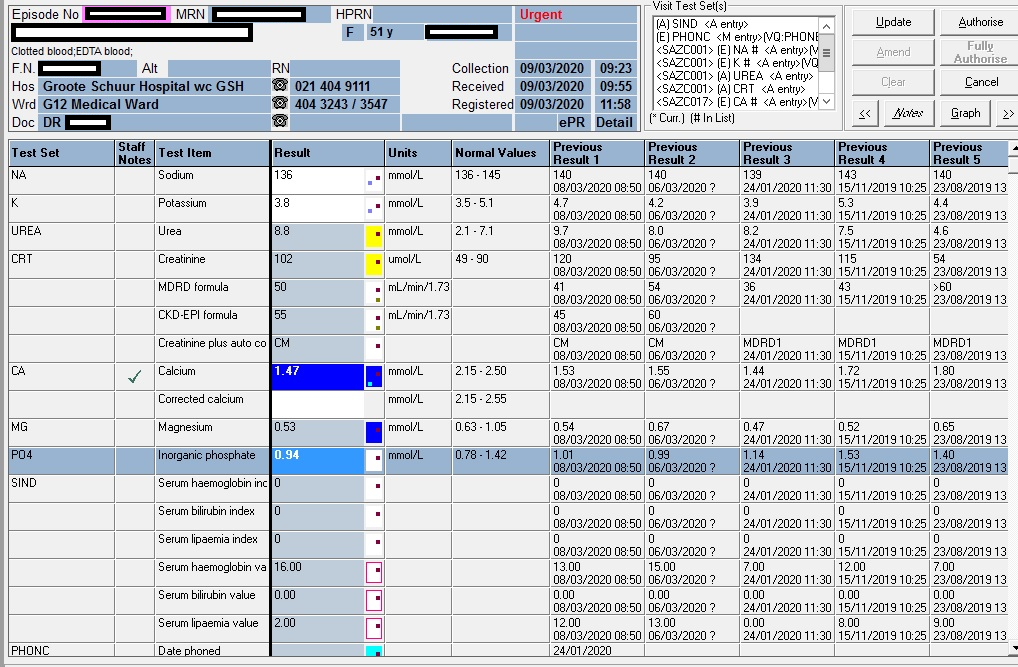
Total calcium of 1.47 mmol/L (2.15 – 2.50)

Presenting Complaint

The patient has been having persistent hypocalcemia despite supplementation with calcium.

History

Figure 1 – Illustration of the patient’s CMP over time:
Calcium: blue; Magnesium: orange; Phosphate: grey
Reference ranges are the horizontal lines without dotted markers

Examination

Not available.

The typical findings in a patient with true hypocalcemia (low ionised calcium) are

Trouseau’s sign

Chvostek’s sign

Laboratory Investigations

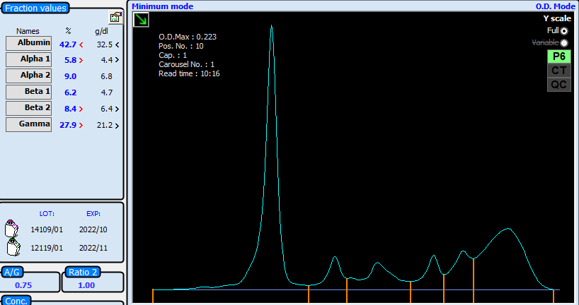
Arguably, the first important consideration in patients with low calcium is the albumin. The patient had a mean albumin of 12 g/L, significantly lower than normal (40-50g/L). Arguably, the calcium can be corrected with the well known Payne’s formula to then be 1.47 + (0.02 x (40-12) = 2.03 mmol/L:

Albumin-adjusted calcium (mmol/L) = total calcium (mmol/L) + 0.02 [40 – albumin (g/L)])

Payne RB, Little AJ, Williams RB, Milner JP. Interpretation of serum calcium in patient with abnormal serum proteins. Br Med J. 1973;4:643-646. DOI: 10.1136/bmj.4.5893.643. (View)

Measurement of serum intact parathyroid hormone (PTH) should be performed in all patients with hypocalcemia; it is the most valuable laboratory test for determining the etiology of hypocalcemia:

  2019/11/15 2019/06/28 2018/08/03
PTH (pmol/L)  21,8 H  15,5 H  25,8 H
Reference interval: (1.6-6.9 pmol/L)

Vitamin D

09/09/2020 15/11/2019 03/08/2018
Total Vitamin D (25-OH VitD) 20.5 nmol/L 45.4 nmol/L 23.2 nmol/L
Guidelines for assessment of Vitamin D status:
<30 nmol/L <12 ng/mL Deficient
30-50 nmol/L 12-20 ng/mL Insufficient
>50 nmol/L >20 ng/mL Sufficient
125-150 nmol/L 50-60 ng/mL Safe upper limit
Reference: Revised South African Clinical Guideline for the diagnosis and management of osteoporosis (NOFSA 2017), endorsing the institute of Medicine Dietary Reference intakes for calcium and vitamin D (2010). Note regarding conversion of units:
Divide result in nmol/L by 2.496 to convert to ng/mL
Multiply result in ng/mL by 2.496 to convert to nmol/L

Other Investigations

Anti-Tissue Transglutaminase antibodies: Negative: repeated 3 months apart, with sufficient IgA levels in the serum): 0.9 & 0.8 U/mL (EliA c/o: 6.9)

Anti-Gliadin antibodies: Equivocal: 7.8 & 9.6 U/mL (EliA c/o: 6.9)

Anti-endomysial antibodies: Negative

HLA-DQ2: Positive

HLA-DQ8: Negative

Final Diagnosis

Hypocalcemia likely due to malabsorbtion (telangiectasia stated by the clinicians).

Take Home Message

According to International guidelines the following association is expected for patients with Coeliac Disease:
Positive for HLA-DQ2 (HLA-DQA1*05, DQB1*02)
Positive for HLA-DQ8 (HLA-DQA1*03, DQB1*03:02)

Considering the fact that the albumin was high with an increased PTH, the calcium very likely was physiologically also low (bioactive Ca). The Payne’s formula also failed to correct the calcium to the normal reference range.


The Vitamin D conundrum

HOSP # WARD F22 Orthopaedics Ward
CONSULTANT   Dr. Jody Rusch DOB/AGE 42 Y Female

Abnormal Result

Total Vitamin D of 27.1 nmol/L on 18 March 2020.

Total Vitamin D of 65.4 nmol/L on 01 April 2020.

Presenting Complaint

Patient had a low impact femur fracture on 18 March 2020 :

History

Patient is known with:

  • previous deep venous thrombosis in 2018, on Warfarin therapy
  • ?Epilepsy, patient is on carbamazepine, for which the Endocrinology specialists were of opinion that it may have been the cause of the low Vitamin D level.

Examination

Laboratory Investigations

Serum protein electrophoresis pattern in keeping with an inflammatory process

Other Investigations

DVT in 2018

Final Diagnosis

Vitamin D deficiency likely due to carbamazepine therapy.

Take Home Message

I was not aware that patients on carbamazepine (or other enzyme inducing agents) have lower Vitamin D levels, and it became evident after a quick literature search that it was in fact the case, see the abstract of the article below, also see another article written by a colleague of mine, Jusine Cole, on the Vitamin D controversies.

Also, I have learned that although “total Vitamin D”, as the assay is named in our immunoassay package insert and on TrakCare LIS, has to do with the total portion with regards to protein binding (to Vitamin D binding protein) and not so much to the fact that calcitriol and calcidiol is measured.

It is however evident that, since the cross-reaction in the immuno-assay is quite pronounced with the various forms of Vitamin D, that total indeed, might be an accurate description. In reality, the assay is however called the Total 25-hydroxy Vitamin D.

The Roche Package insert values for specificity for the Total 25-OH Vitamin D assay.

Vitamin D levels and bone turnover in epilepsy patients taking carbamazepine or oxcarbazepine.

Abstract

PURPOSE:

Evidence suggests that enzyme-inducing antiepileptic drugs (AEDs) may decrease serum 25-hydroxyvitamin D (25-OHD) levels and increase bone turnover. We sought to determine whether these are affected by treatment with carbamazepine (CBZ) or oxcarbazepine (OXC).

METHODS:

We measured serum levels of 25-OHD, parathyroid hormone (PTH), osteocalcin (OCLN), bone alkaline phosphatase (BAP), and urinary N-telopeptides of type I collagen cross-links (NTX) in normal controls (n=24) and in epilepsy patients taking CBZ (n=21) or OXC (n=24) in monotherapy. CBZ patients were subsequently switched overnight to OXC monotherapy, and after 6 weeks, the tests were repeated.

RESULTS:

25-OHD levels were lower in each drug-treated group (OXC, 19.4+/-2.3 pg/ml; CBZ, 20.4+/-2.4) than in the controls (27.5+/-2.8) (ANOVA, p=0.052). This difference was significant for the OXC group (p<0.05). PTH, BAP, and NTX did not differ significantly among groups. OCLN levels were somewhat elevated in the OXC group (2.79+/-0.47 ng/ml) and more clearly and significantly elevated in the CBZ group (3.63+/-0.36) compared with controls (2.38+/- 0.41) (p=0.053). Because the data were very similar between OXC and CBZ groups, they were combined to increase statistical power. The combined drug-treatment group had significantly higher BAP (p=0.02) and lower 25-OHD (p=0.015) than did controls. The latter remained significant even after accounting for the confounding effects of age on 25-OHD levels (p<0.05). No significant differences were found after CBZ patients were switched to OXC.

CONCLUSIONS:

Epilepsy patients taking OXC or CBZ have significantly lower 25-OHD than do normal controls, with a pattern of changes in other bone biomarkers suggestive of secondary hyperparathyroidism. It may be prudent for patients taking CBZ or OXC to be prescribed 25-OHD replacement.